Cirugía de Columna y articulaciones

Cirugía de Columna y articulaciones
Cirugía de Columna y Articulaciones

domingo, 9 de febrero de 2014

Stem cell injections improve spinal injuries in rats

http://www.kurzweilai.net/stem-cell-injections-improve-spinal-injuries-in-rats

Stem cell injections improve spinal injuries in rats 

May 29, 2013
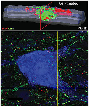
A three-dimensional, reconstructed MRI image (upper) shows a cavity caused by a spinal injury nearly filled with grafted neural stem cells, colored green. The lower image depicts neuronal outgrowth from transplanted human neurons (green) and development of putative contacts (yellow dots) with host neurons (blue). (Credit: UC San Diego School of Medicine) 
A single injection of human neural stem cells produced neuronal regeneration and improvement of function and mobility in rats impaired by an acute spinal cord injury (SCI), an international team led by researchers at the University of California, San Diego School of Medicine reports
Grafting neural stem cells derived from a human fetal spinal cord to the rats’ spinal injury site produced an array of therapeutic benefits — from less muscle spasticity to new connections between the injected stem cells and surviving host neurons.
“The primary benefits were improvement in the positioning and control of paws during walking tests and suppression of muscle spasticity,” said Martin Marsala, MD, professor in the Department of Anesthesiology and a specialist in spinal cord trauma and spinal injury-related disorders.
Spasticity — exaggerated muscle tone or uncontrolled spasms — is a serious and common complication of traumatic injury to the spinal cord.
“In all cell-grafted animals, there was robust engraftment, and neuronal maturation of grafted human neurons was noted,” Marsala said. “Importantly, cysts or cavities that can form in or around spinal injuries were not present in any cell-treated animal. The injury-caused cavity was completely filled by grafted cells.”
The rats received the pure stem cell grafts three days after injury (no other supporting materials were used) and were given drugs to suppress an immune response to the foreign stem cells. Marsala said grafting at any time after the injury appears likely to work in terms of blocking the formation of spinal injury cavities, but that more work would be required to determine how timing affects functional neurological benefit.
The grafted stem cells, according to Marsala, appear to be doing two things: stimulating host neuron regeneration and partially replacing the function of lost neurons.
“Grafted spinal stem cells are rich source of different growth factors which can have a neuroprotective effect and can promote sprouting of nerve fibers of the host neurons,” he said. “We have also demonstrated that grafted neurons can develop contacts with the host neurons and, to some extent, restore the connectivity between centers, above and below the injury, which are involved in motor and sensory processing.”
The scientists used a line of human embryonic stem cells recently approved for Phase 1 human trials in patients with chronic traumatic spinal injuries. Marsala said the ultimate goal is to develop neural precursor cells (capable of becoming any of the three main cell types in the nervous system) from induced pluripotent stem cells derived from patients, which would likely eliminate the need for immunosuppression treatment.
Human trials
Pending approval by UC San Diego’s Institutional Review Board, the next step is a small phase 1 trial to test safety and efficacy with patients who have suffered a thoracic spinal cord injury (between vertebrae T2-T12) one to two years earlier, and who have no motor or sensory function at or below the spinal injury site.
“This is exciting, especially because, historically, there has been very little to offer patients with acute spinal cord injury,” said study co-author Joseph Ciacci, MD, professor of surgery and program director of the Neurosurgery Residency at the UC San Diego School of Medicine. Ciacci, who is also chief of neurosurgery for the Veterans Affairs San Diego Healthcare System, will oversee the clinical trial at UC San Diego and the VA.
Ciacci said if the initial study confirms safety and efficacy, as well as the viability of the implanted cells, neural regeneration and decreased spasticity, the protocol can be expanded to other patients with other forms of severe spinal cord injury.
Colleagues at UC San Diego and in Slovakia, the Czech Republic, and The Netherlands, were also involved in the research.
Funding came, in part, from the California Institute for Regenerative Medicine and Neuralstem Inc., which also provided cells.
Topics: Biomed/Longevity | Cognitive Science/Neuroscience

~11% of Combat Wounded Have Spinal Injuries

http://www.empr.com/11-of-combat-wounded-have-spinal-injuries/article/312216/


~11% of Combat Wounded Have Spinal Injuries 

~11 Percent of Combat Wounded Have Spinal Injuries
(HealthDay News) – Spine injuries account for >11% of all casualties among U.S. combat-wounded military in Iraq and Afghanistan, according to a study published in the Sept. 15 issue of Spine.
Andrew J. Schoenfeld, MD, from the Texas Tech University Health Sciences Center in El Paso, and colleagues conducted a retrospective analysis of casualty records from the Department of Defense Trauma Registry for 2005–2009 to assess the incidence and epidemiology of combat-related spinal injuries for the wars in Afghanistan and Iraq.
The researchers identified 872 casualties with spine injuries among a total of 7,877 combat wounded (11.1%), with a mean age of 26.6 years. The most common injury morphology was spine fractures, accounting for 83% of all spinal wounds. The incidence of combat-related spinal trauma was 4.4 per 10,000, while spine fracture incidence was 4 per 10,000. The rate of spinal cord injuries was 4 per 100,000. The incidence of spinal cord injuries was increased in Afghanistan (incidence rate ratio [IRR], 1.96), among Army personnel (IRR, 16.85), and in 2007 (IRR, 1.9). Gunshot-related spinal injuries were significantly more likely to occur in Iraq than Afghanistan (17% vs. 10%; P=0.02).
"The incidence of spine trauma in modern warfare exceeds reported rates from earlier conflicts," the authors write.
Relevant financial activities outside the submitted work were disclosed.

 2013 Sep 15;38(20):1770-8. doi: 10.1097/BRS.0b013e31829ef226.

Spinal injuries in United States military personnel deployed to Iraq and Afghanistan: an epidemiological investigation involving 7877 combat casualties from 2005 to 2009.

Abstract

STUDY DESIGN:

Retrospective analysis of a prospective data set.

OBJECTIVE:

Determine the incidence and epidemiology of combat-related spinal injuries for the wars in Afghanistan and Iraq.

SUMMARY OF BACKGROUND DATA:

Recent studies have identified a marked increase in the rate of combat-related spine trauma among casualties in Afghanistan and Iraq. Limitations in these previous works, however, limit their capacity for generalization.

METHODS:

A manual search of casualty records stored in the Department of Defense Trauma Registry was performed for the years 2005 to 2009. Demographic information, nature of spinal wounding, injury mechanism, concomitant injuries, year, and location of injury were recorded for all soldiers identified as having sustained combat-related spine trauma. Incidence rates were constructed by comparing the frequencies of spine casualties against defense manpower deployment data. Multivariate Poisson regression was used to identify statistically significant factors associated with spinal injury.

RESULTS:

In the years 2005 to 2009, 872 (11.1%) casualties with spine injuries were identified among a total of 7877 combat wounded. The mean age of spine casualties was 26.6 years. Spine fractures were the most common injury morphology, comprising 83% of all spinal wounds. The incidence of combat-related spinal trauma was 4.4 per 10,000, whereas that of spine fractures was 4.0 per 10,000. Spinal cord injuries occurred at a rate of 4.0 per 100,000. Spinal cord injuries were most likely to occur in Afghanistan (incident rate ratio: 1.96; 95% confidence interval: 1.68-2.28), among Army personnel (incident rate ratio: 16.85; 95% confidence interval: 8.39-33.84), and in the year 2007 (incident rate ratio: 1.90; 95% confidence interval: 1.55-2.32). Spinal injuries from gunshot were significantly more likely to occur in Iraq (17%) than in Afghanistan (10%, P = 0.02).

CONCLUSION:

The incidence of spine trauma in modern warfare exceeds reported rates from earlier conflicts. The study design and population size may enhance the capacity for generalization of our findings.

LEVEL OF EVIDENCE:

3.
PMID:
 
23759821
 
[PubMed - in process]

Proof That Functional Strength Training Cures Low Back Problems and Pilates Won't

http://breakingmuscle.com/mobility-recovery/proof-that-functional-strength-training-cures-low-back-problems-and-pilates-wont

Proof That Functional Strength Training Cures Low Back Problems and Pilates Won't 

The following is a guest post by Andrew Lock:
Mention low back rehabilitation to people who believe in Pilates and you will tend to get that same faraway look in their glazed eyes that I see in the eyes of people who attend crystal healing workshops and who whisper about the alien anal probes they were subjected to when they were abducted by the little green men (in their reformer bed spaceships, no doubt).
So I am here to give to you, the trainers in the trenches, just what you need to have in your armory when engaging with one of these navel pullers of the New Age. Rather than smacking them upside the head with the rolled-up weekend edition of your local newspaper, as I usually do, you can now roll up a copy of this article and stuff it deep into their incredulous, slack-jawed mouths.
To begin de-programming a Pilates cult member, I suggest you take a leaf fromA Clockwork Orange and strap the brainwashed subject into a barber’s chair, fix back their eyelids, and show them these MRI scans. If a picture says a thousand words, these pictures say a hell of a lot more:
spine anatomy, spine stabilization, pilates, pilates lies, truth about pilates
Left: Pilates result; Right: Functional strength rehab result
The two MRI scans you see here are clear, undeniable proof of the failure of long-term Pilates training and the success of functional strength rehabilitation methods to cure low back pain.
The MRI on the left is from a person who came to see me about her years ofunresolved low back pain . She had been doing “clinical Pilates” three to four times per week for the last four years, and was still having low back pain.What you see in her scan is atrophic multifidus muscle in the lumbar spine. This is after four years of so-called rehabilitation. Her spinal musculature is lacking stabilizing muscle and is fat infiltrated. Her scan is otherwise clear of any problems. There is no disc injury, just poor musculature, and she was doing Pilates at one of the most respected Pilates teaching and rehabilitation centers in the country. And yes, she could perform an isolated transverse abdominis contraction .
The scan on the right is a guy who came to see me who had experienced a huge disc injury, including nerve involvement. Did that worry me? No. I’ve treated over 10,000 patients in the last twenty years and I began weight training as an athlete before I was a physical therapist. I assessed him and began him on functional strength rehabilitation. What you see in his scan is the result of his rehab. Massive paraspinal musculature and maximum multifidus hypertrophy.
What did he do for rehab? He did exactly what I prescribe - a progressive protocol that included deadlifting (he knew how to do it) in the early stage and culminated with double kettlebell swings and kettlebell Sots pressing at the end stage of rehab. The exact protocol I prescribe varies upon the patient’s presentation, but the paradigm is clear - progressive overload and functional movement loading. This athlete continued in his training, even with a bulge (I taught him the skills to manage the problem in training, I’ll teach you that in another article) and competed successfully at an international level weeks after the scan was taken.
Let’s take a closer look at the scans now so you can really sink your teeth into the Pilates jugular. I’ll start with what resulted from four years of clinical Pilates and transverse abdominis isolation rehabilitation protocols.
You don’t need a degree in radiology to understand these MRI scans, but I’ve helped to define the points I think are best. I’m not a graphic artist, just a genius, so put up with my efforts - it’s worth it.
spine anatomy, spine stabilization, pilates, pilates lies, truth about pilates
In this scan, the area where the fat is sitting is where the deeper layers of the multifidus muscle should be. It is not there. Fat is, and fat is not a very functional tissue to have next to your spine. The muscle you see is the dark tissues as indicated and the bone has been outlined for clarification. What you see is the result of non-functional transverse abdominis focused “core” training.Though they used the term “functional core,” it failed that requirement spectacularly. Why have these methods failed this person, and why has it been failing athletes as a method of low back rehabilitation? Move over and let me drive, I understand these streets.
First, let’s begin by enjoying the fruits of our intellectual labors, and revel in the results of functional strength rehabilitation.
spine anatomy, spine stabilization, pilates, pilates lies, truth about pilates
Muscle, muscle everywhere! Look at this scan it could come from a damn rodeo bull! Everything that should be hypertrophied is. This was after just four months of functional strength rehab. There is no mystery as to why functional strength rehabilitation protocols work and why core stability and Pilates methods fail. It is all in the science of anatomy and movement.
I have been studying science, injury, rehabilitation and strength for about 25 years. This involved thousands of patients and a lot of post-graduate study. I cannot show you, today, the exact methods I use to assess and rehab in this short article. This article is about results and the obvious science of successful rehabilitation principles. Principles you already know. I’m going to now put in place some of the foundation stones of successful rehabilitation, and highlight how the Pilates and core approach fails those basic human physiological principles.
Key points to be addressed in the low back rehabilitation:
  • Consequent to low pain injury the multifidus muscle of the spine atrophies.
  • People with low back injury have altered movement patterns after injury.
  • Over 90% of people who injure their low back will have more episodes of injury.
So let us look at the facts of functional strength rehabilitation:
SAID = Specific Adaptation to Imposed Demand.
Yes, you all knew that. But why has it not been applied in the field of lumbar spine rehabilitation?
You must produce hypertrophy of the lumbar multifidus muscle. How does “pulling your bellybutton in towards your spine” while lying upon a reformer bed waving your arms and legs about like a dying turtle on its back in the midday sun produce a hypertrophic adaptation? Uh, it doesn’t, Beavis. The Pilates methods fail to impose significant enough overload to the musculature to produce functional hypertrophy. There is no overload to produce muscular hypertrophy that will translate to athletic demands. The specific adaptation you learn from Pilates is specific only to Pilates.
spine anatomy, spine stabilization, pilates, pilates lies, truth about pilates
Functional rehabilitation requires movement specific overload, e.g. deadlift, kettlebell swing. Spectacularly, the research parroted by these transverse abdominis advocates is a study by Hoffer and Andreasson that contends contractions as low as 25% maximum voluntary contraction are able to provide maximal joint stiffness. This reference was a study of the soleus muscle of a cat that had been decerebrated (removed the cerebral brain function from). Do you think the study of a soleus muscle in a cat (quadruped), which had been brain injured, is reasonable support, or applicable to human beings (biped without brain injury)? But these numbers are often quoted by supporters of Pilates. 25% of a maximum contraction of transverse abdominis will not result in overload and hypertrophy of the multifidus, which is required for rehab. Also, transverse abdominis is not a lumbar stabilizing co-contractor with multifidus. No, it is not. Never was, and never will be.
Altered movement patterns are noted consequent to low back injury. Yes, transverse abdominis is supposed to be the first muscle to contract in human movement, but what is the best way to restore normal movement patterns (including restoring transverse abdominis patterning)? It’s not by lying on your back or being on your hands and knees.Restore normal movement patterns by learning normal movement patterns, not abnormal ones.
So teaching a person to move correctly and to control a lumbar lordosis under load produces the single most effective method of overload, movement specific function, and pain-free low back restoration. You begindeadlifting and kettlebell swinging early. This keys into why back pain is recurrent. People are not addressing functional movement patterns. By functional movement pattern I do not mean that confounding, stupid fitball exercise involving some sort of damn cable machine and a delirious rapturous grin from a personal trainer. The rule of thumb these people seem to use is, “If it looks dangerous or stupid, tell the client it is for their functional core.” In science functional means functional, not fucking stupid.
Now, there are important protocols, assessments and progressions within functional strength rehabilitation to produce the outcome I have demonstrated.This article is not here to show you what they are. Those articles come later.This article is specifically focused to show the superiority of progressive overload and applied strength science to the superstition and blind belief systems that now dominate low back rehab in the minds many health professionals. It is time for professional trainers to take back the rehabilitation work that is really theirs in the first place. It might mean a lot more study for you, but I’ll also be the first to tell you - being smart feels good.
Okay, now I’m off to write a book on the science and methods of successful strength rehabilitation. Now let’s see, how will I begin? I know I’ll begin with, “Call me Ishmael.”
References:
1. Hoffer J Andreasson S 1981 Regulation of soleus muscle stiffness in premammillary cats . Journal of Neurophysiology 45: 267-285
MRI scans property of Andrew Lock.
Cat photo courtesy of Shutterstock .

‘Watch’ issued about femoral neck fractures in long-necked modular implants

http://www.healio.com/orthopedics/hip/news/online/%7B0c8a4d00-a3ac-4482-8fd5-a1e6242252d8%7D/watch-issued-about-femoral-neck-fractures-in-long-necked-modular-implants


‘Watch’ issued about femoral neck fractures in long-necked modular implants 

  • January 31, 2014
JBJS Case Connector has issued a “Watch” regarding femoral neck fractures in patients whose implants used modular head-neck and neck-stem designs. While some of the designs are no longer available from manufacturers, such devices have been implanted and the “Watch” encourages surgeons to be wary about long femoral necks in the modular hip designs.
The “Watch” is based on a case presented in the Jan. 22 issue of JBJS Case Connector and three previously published cases which all cite modularity and long femoral neck length as common denominators of a risk of fracture that should be discussed with patients.
“The ‘Watch’ designation may encourage the orthopedic community to either demonstrate that these are isolated, unrelated cases or sharpen the focus further by rigorously evaluating the intervention and reporting related cases,”Marc Swiontkowski, MD, editor of JBJS Case Connector, stated in a press release.
Reference:
Swiontkowski M. JBJS Case Connector. 2014;doi:10.2106/JBJS.CC.M.00301.
Disclosure: Swiontkowski received consulting fees from Eli Lilly, METRC and Pfizer China, participation fees from Eli Lilly and METRC and royalties from Lippincott. He is a consultant for Eli Lilly, employed by JBJS and receives royalties from Lippincott.